Medical image processing using machine learning (ML), significantly deep learning (DL) shows specific trust in tasks from tumor prediction to organ partition. In this area we always hold Convolutional Neural Networks (CNNs) because of their ability to Automate and adjust learning dimensional hierarchies of features from images.
Below are the constructing steps that we implement in medical image processing using ML:
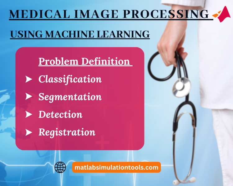
- Problem Definition:
State the particular task:
- Classification: We examine the type of disease and its symptoms from an image.
- Segmentation: Describe state of interests in an image of our project.
- Detection: Tumors like abnormalities are the particular features we find in the image.
- Registration: We organize various sets of pictures.
- Data Collection:
- It is efficient for us to utilize source labeled medical images from the public datasets such as National Institutes of Health (NIH) datasets, Kaggle limitations and coordinating with medical institutions.
- To make sure the security of the patient, we hide and de-identify the data.
- Pre-processing the Data:
- Image Resizing: To ensure every image is in continuous size we do image cropping.
- Normalization: We measure pixel values range [0-1] frequently.
- Data Augmentation: By using conversions such as rotations, translations and flips we artificially raise the size of the dataset and enhance model productivity.
- Exploratory Data Analysis (EDA):
- We visualize sample images and their related labels.
- Analyzing the dispersions of various classes and conditions in our project.
- Detecting class imbalance assists us in work.
- Feature Engineering (for traditional ML):
- Texture Analysis: By retrieving texture descriptors we determine the data.
- Shape Descriptors: To catch the shape of organs and anomalies we create features.
- Statistical Features: Mean, variance and other statistical scales of pixel values are properties we use in this process.
- In particular, we utilize CNNs for DL to extract features and inherit the structure.
- Model Selection:
- Existing ML: SVM, Random Forests are the traditional ML models we implement.
- DL: We incorporate CNNs such as VGG, ResNet, and U-Net for segmentation and transfer learning by pre-trained models are useful because of insufficient medical image data.
- Training the Model:
- By dividing the data we perform training, evaluation and validation sets.
- To prevent overfitting we train the framework on the instructing dataset.
- Evaluation:
- Accuracy: We scale the proportion of appropriate classifications.
- Dice Coefficient: In general for segmentation tasks we scale the overlap between the detected and real partitions.
- Sensitivity, Specificity: It is essential to the medical context where false negatives and false positives have certain suggestions in our project.
- ROC-AUC: For binary classification tasks we use this technique.
- Optimization:
- Adjust our model with hyperparameters.
- We examine grouping approaches and model integrations.
- Deployment:
- Based on the usage we apply our system in a medical setting and make sure in real-time and periodic processing when needed.
- Feedback Loop:
- In real-world situations we consistently track our system’s efficiency.
- We collect reviews from professionals and combine it into subsequent repetitions.
Tools & Libraries:
- Data Handling & EDA: Pandas, NumPy, Matplotlib and Seaborn support our project.
- Image Processing: OpenCV and SimpleITK are helpful for us in processing an image.
- Modeling: We implement scikit-learn, TensorFlow, Keras and PyTorch.
- Segmentation-specific: U-Net structures are valuable for our model.
Ethical & Practical Considerations:
- Data Privacy: We make sure permissions to privacy standards like HIPAA in the US.
- Model Interpretability: Grad-CAM is beneficial for our project when the challenging nature of medical decisions, model understandability is crucial.
- Collaboration: It is essential for us to consult with clinical experts who are skilled in ML. By this we achieve better solutions and reliable models.
In overview, medical image processing using ML contains huge possibilities for reforming healthcare. By using the appropriate technique and integration we serve in diagnosis, treatment planning and medical research with our model.
Medical Image Processing Using Machine Learning Thesis Ideas
Dive deep into crucial tips and gain valuable perspectives for crafting your Medical Image Processing Using Machine Learning Thesis Concepts. Stay updated with our cutting-edge concepts from our specialists. We prioritize punctuality, ensuring project completion well within the deadline. Interested in discovering how matlabsimulation.com consultancy services can elevate your academic performance? Schedule a consultation with our advisor via email, Google Meet, or phone call! Our proficient team is here to address your inquiries and assist you in making well-informed choices for your academic pursuit. Explore some of the thesis idea we have shared in this page.
- Skin Cancer Detection and Classification System by Applying Image Processing and Machine Learning Techniques
Keywords
Deep learning, skin cancer, convolutional network
A major aim of this article is to detect the skin lesion and to categorize it as benign or malignant. Our suggest article includes preprocessing of images like noise reduction, segmentation, and feature extraction from the lesion parts. After that, feature selection process is carried to precisely label the lesion part. In this, SVM classifier is utilized to identify the skin lesion as benign or malignant.
- A Transfer Learning Based Classification of Nephrolithiasis Using CT Scan Images Employing Machine Learning with Image Processing
Keywords
Kidney stone, CT scan, Artificial intelligence, Multilayer perceptron, Histogram equalization, FPGA
By using AI, early forecasting of kidney stones is carried out using CT scan. To categorize the kidney stone, backpropagation network with multilayer perceptron is employed. We performed segmentation process in two phases. Firstly, kidney is segmented. Secondly, stone is precisely segmented. Then the preprocessing steps are carried out and improved using histogram equalization. At last, CADx is employed as a tool for identification and diagnosis.
- Machine Learning for Medical Image Analysis: A Survey
Keywords
Medical Imaging, Compression, Detection
Several ML and DL approaches are reviewed in our paper that are commonly utilized for image preprocessing steps like image identification, segmentation of images, categorization and image compression using different images such as CT, MRI and x-ray radiography. For identification and categorization of various diseases, CNN provides greater efficiency in autonomous feature extraction procedure.
- Cancer Detection Using Image Processing and Machine Learning
Keywords
Image-processing, Anisotropic Filtering, Watershed algorithm, Thresholding
Image processing procedure assists the radiologists in detecting various diseases. It is an efficient approach to examine the image and suggest the outcomes. This study suggested a model to identify various cancers such as Blood Cancer, Brain Tumor, Breast Cancer and Lung cancer. Several approaches are used to identify cancers like segmentation procedure, morphological operations and some method is also utilized like watershed segmentation method.
- Machine learning based biomedical image processing for echocardiographic images
Keywords
Biomedical imaging, Image classification, Image segmentation, Machine learning algorithms, neural networks, Regression analysis
In our paper, K-Nearest Neighbor (KNN) is utilized for image segmentation process. Extraction of images and categorization of data are carried out related to neural networks. We stated that, KNN approach is very user friendly and efficient that is mostly used for image processing procedures like segmentation, extraction and classification. Gray level co-occurrence matrix features are utilized in our suggested framework.
- Machine Learning in Medical Image Processing
Keywords
Feature extraction, Cancer classification, Histopathological images, HI, Magnetic resonance imaging (MRI), Mammogram images, Supervised ML, Unsupervised ML
Several ML approaches are utilized for medical image processing is reviewed in our paper. This review mainly concentrated on the categorization of medical images of several sections of human body. Categorization of diseases as tumor, non-tumor or other categories is carried out using different methods. Various procedures such as segmentation and feature extraction are also discussed in our paper.
- Machine Learning-Based Categorization of Brain Tumor Using Image Processing
Keywords
Merciless tumor, benign tumor, Backpropagation, Otsu thresholding, k-means clustering
Edge-based Contourlet Transformation is involved as a main step in our recommended framework for registration procedure. Tumor point segmentation is performed in our paper. After that, extraction of features is carried out, in that, different features such as Otsu’s thresholding, k-means are integrated and local binary marking texture is utilized for identification of meningioma. Finally, categorization process is performed based on neural network technique.
- Detection of Malaria Disease Using Image Processing and Machine Learning.
Keywords
Malaria disease, Blood smear images, Computer-aided diagnosis
By employing various ML approaches and image processing techniques, a computer-aided approach is developed for autonomous identification of malaria parasites. Features are extracted from the red blood cell images to categorize the blood cells as parasitized or uninfected. Several ML classifiers are utilized such as AdaBoost, K-Nearest Neighbor, Decision Tree, Random Forest, Support Vector Machine and Multinomial Naive Bayes.
- Machine Learning and Digital Image Processing in Lung Cancer Detection
Keywords
Lung Cancer, Deep CNN, ResNetv101, VGG-16
In lung cancer detection, several procedures are followed in our paper such as preprocessing of images, edge detection and segmentation process. These images are utilized by various ML techniques like Deep CNN, ResNetv101, and VGG-16 for the comparison of malignancy categorization. As a consequence, Deep CNN provides greater end results than ResNet101 and VGG-16.
- Identification and Counting of Blood Cells Using Machine Learning and Image Processing
Keywords
Blood cells, Chan vase, S-CNN
In our study, different kinds of blood cells such as Red blood cells (RBC), white blood cells (WBC), and platelets are autonomously identified and counted by utilizing the procedure of image segmentation and ML techniques like S-CNN (Suit-Convolutional Neural Network). By using images of character assassination, blood cells are counted in a minimum amount of time through computer-aided monitoring and detection approach.